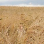

Fusarium head blight and the mycotoxins it cause, especially deoxynivalenol (DON), can be a serious economic detriment to producers. It reduces yield and grain grade, as well as contaminates the grain, making it dangerous for human and animal consumption.
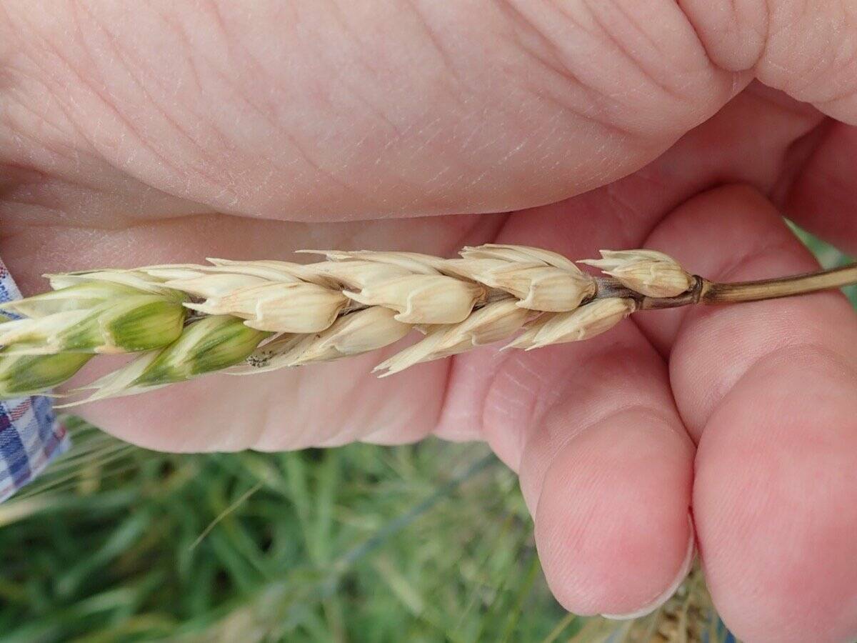
Infected wheat kernels, known as fusarium-damaged kernels (FDK), can be visually assessed by a producer, typically appearing chalky white, shrunken and sometimes covered in pink or white mycelium. But other cereals aren’t as easily assessed, such as barley, which has a hull that hides the infection.
The visual assessment is also subjective, labour-intensive and misleading, as contaminated kernels can appear healthy.
Read Also
MANITOBA AG DAYS: Don’t wait to buy fertilizer, farmers warned
Higher fertilizer prices likely ahead, says Ag Days speaker. Farmers waiting until spring to buy fertilizer might end up eating the cost.
WHY IT MATTERS: Fusarium infection in cereals can become a serious marketing problem for the farmer.

Sheila Andrade, a PhD student at the University of Saskatchewan, has been working on developing a method of detection to ease the struggles of producers, agronomists and industry.
“DON is measured by chromatography and immunological methods, which is costly, destructive and also time-consuming,” she explained during her presentation at the Pan-American Light Sources for Agriculture conference.
The disease affects kernel development, and both DON and FDK are downgrading factors, with FDK often used as a proxy for DON contamination. However, those correlations are not consistent, since contamination is also influenced by the genotype isolate and the time of kernel infection. Because of this, Andrade has been studying an alternative method of detection.
Andrade is working with what she describes as “a synchrotron-based x-ray phase-contrast computed tomography” — essentially using powerful x-ray imaging at the Canadian Light Source (CLS). Her objective is to measure the physical traits of fusarium-infected wheat kernels, link those traits with DON contamination, and compare how fusarium symptoms differ between durum and bread wheat.
Once these parameters and the images are understood, they can be used to inform machine-learning technology to create an easy disease detection method.
Research process
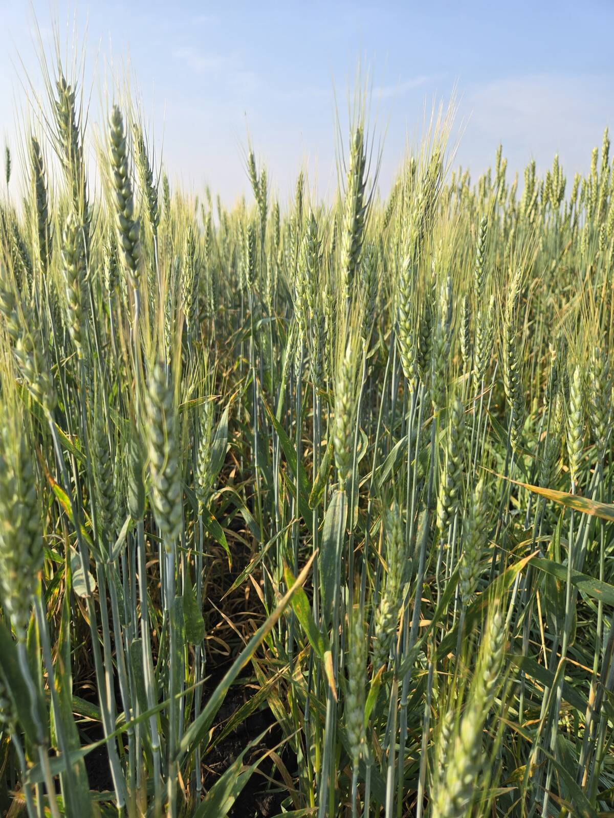
There were four varieties of durum wheat and five varieties of bread wheat varieties used in the research. Each variety was planted in field nurseries and inoculated with Fusarium graminearum at the flowering growth stage. When they reached maturity, they were harvested and then went to CLS for Andrade’s research.
The wheat spikes were placed in test tubes, and scanned in batches of six using the biomedical imaging beamline at CLS. This beamline enabled Andrade and her team to non-destructively view the wheat spike from different angles and digitally reconstruct the kernels in 3D.
From there, the test tubes were separated and individual kernels were isolated. Researchers were then able to measure traits used to identify FDK, including total area, length, width and volume, as well as shape factor, density, grey mass and thickness.
“After we finish all the image processing and the segmentation, we wanted to know how the kernel was contaminated and the level of contamination of each individual kernel,” Andrade said.
However, to determine the contamination, the team had to manually thresh the wheat spikes and label each kernel to its corresponding image analysis. In total, they manually threshed 725 kernels and classified them into two groups: 369 DON-free and 356 DON-contaminated kernels.
This information was analyzed to learn how the parameters would shift between contaminated and non-contaminated. Andrade explained that a significant difference was found between the groups for most parameters, except for length. The DON-free kernels had high values in most traits, except for shape factor, as expected.
There were also significant interactions with the wheat type, where the physical differences were more evident in bread wheat.
Machine-learning model
By using the imaging from the synchrotron beamlines, Andrade determined that average size, density, shape, length and grey mass were the most important features in determining DON-contaminated versus DON-free kernels.
“We have some interesting results showing that, for example, density and gray mass and sometimes shape differ between the infected and non-infected kernels,” she said. “This why we got this significant difference, and why we use those parameters to train a machine-learning model.”
Using this information, Andrade and her colleagues tested four different machine-learning models. They trained the models with 70 per cent of their data set and reserved the remaining 30 per cent to test accuracy. One model performed best, correctly classifying about 80 per cent of DON-contaminated kernels and 82 per cent of DON-free kernels. However, it still missed about 20 per cent of the contaminated kernels.
Going forward
This was a “first step project” for Andrade. Its goal was to learn if X-ray images would be an effective way to separate kernels. So far, it’s been a success.
She said the next goal would be to use a portable machine to benefit wheat breeders, who have a lot of materials to analyze, and could eventually be used by agronomists, farmers and the industry.
A machine of this sort would be a game-changer in disease detection, helping better identify DON-contaminated kernels that may slip beneath the radar and cause a potential food security issues.
Andrade hopes that such a machine would be available in the next three to five years, based on machines similar to those used in the lab right now. The team already works closely with members of the engineering department, who developed a lab-level, portable RGB camera. She said she’d like to have something similar but for X-ray.
Potentially, there may be a need for combining more than one image type, such as X-ray and conventional photographic images, since colour can be important for identifying FDK.
Another part of the project is expanding to other cereals. Andrade has recently begun imaging and analyzing barley and will move on to other crops once that work is complete. Barley is a priority since the hull hides infection, leaving breeders, producers and industry “blind.”
“They don’t know what’s happening, so they need to test,” she said. “If we can use the x-ray image to predict DON, it will be really useful for them, because they don’t have the visual assessment to guide them.”
The method is proving helpful for wheat, and Andrade believes it could be even more valuable for barley. Once the key parameters are clear, much of the process can be applied to other cereals. Those should be quicker to image and segment than wheat, though still time-consuming. For now, Andrade has 1,500 samples — each with 100 seeds — from the past three years still waiting to be analyzed.